Ultrasonic Imaging
Core Director: Charles S. Chung, Ph.D.
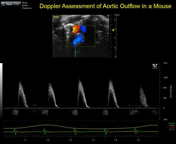
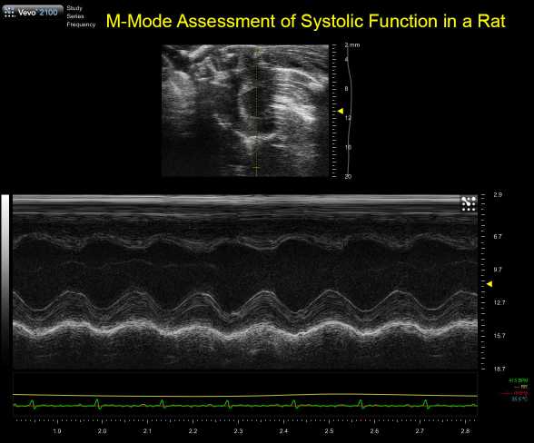
The Department of Physiology maintains a VisualSonics Vevo2100 Small Animal Ultrasound System. Probes suitable for imaging of cardiovascular function and anatomical structures in mice (40MHz center frequency) and rats (21MHz center frequency) are available. The lower frequency probe can also be used to image high velocity constrictions (for Transverse Aortic Constriction models in mice. Heated physiologic monitoring stages (ECG, respiratory rate, temperature) are used for imaging isoflurane anesthetized mice.
Standard echocardiographic measurements include long-axis B-mode, short-axis B-mode, short-axis M-mode, and aortic flow (ascending and arch). Diastolic function including transmitral Doppler and Tissue Doppler Imaging are available, along with right ventricular functional measurements. Advanced measurements including coronary and pulmonary vein flow are also available. Vascular measures, including vessel areas and flow are also available, along with quantification using VisualSonics VevoVasc software. Core staff have experience in ultrasound imaging of diaphragm functional and of the liver and kidney anatomy. Conscious echocardiography is also available to evaluate in vivo contractility at physiologic heart rates.
Core services are generally available at no cost to Primary Physiology Faculty. Contact the Core Director for a consultation and to discuss availability.