Departmental Core Facilities
- Atomic Force Microscopic Imaging
Core Director: Bhanu Jena, Ph.D.
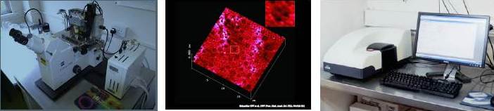
Nano scale molecular and cellular physiology is an important field of Nano Science and Nano Medicine utilizing cutting edge technologies such as atomic force microscopy (AFM) and photon correlation spectroscopy using the Zeta Sizer. Utilizing these technologies, imaging and studying single molecular interactions and their dynamics can be accomplished. Figure below shows the AFM (far left), an image of the surface topology of live acinar cell apical plasma membrane (middle), and the Zeta Sizer (right). These instruments have frequently been used in collaborative studies both by faculty within the department of physiology and from other departments at Wayne State University.
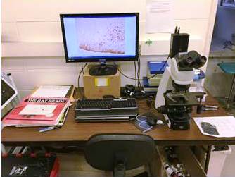
- Confocal Microscopic Imaging
The confocal microscope is a customized workstation for microscopy image acquisition and analysis. This microscope is an inverted Leica SD600 microscope with 5X, 10X, 40X, and 63X oil objectives. The microscope is equipped with an X-Light V1 spinning disk confocal imager (Crest Optics) with automatic 3-position filter wheel. The system is equipped with a laser light source (Multiline Laser Illuminator, LDI-7, 89 North). The LDI-7 generates 7 distinct laser lines with up to 1 W power per line for efficient excitation. The acquisition and image analysis software (MetaMorph NX) is capable of multi-dimensional analysis, compatible with the time and Z-dependent acquisition functions of a spinning disk system. Graphic files can be sent to the Departmental server for individual data storage through a direct internet connection in the room.
Core Director: Alyson Wessells
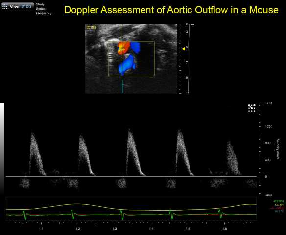
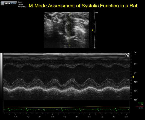
- Ultrasound Imaging
Core Director: Charles S. Chung, Ph.D.
The Department of Physiology maintains a VisualSonics Vevo2100 Small Animal Ultrasound System. Probes suitable for imaging of cardiovascular function and anatomical structures in mice (40MHz center frequency) and rats (21MHz center frequency) are available. The lower frequency probe can also be used to image high velocity constrictions (for Transverse Aortic Constriction models in mice. Heated physiologic monitoring stages (ECG, respiratory rate, temperature) are used for imaging isoflurane anesthetized mice.
Standard echocardiographic measurements include long-axis B-mode, short-axis B-mode, short-axis M-mode, and aortic flow (ascending and arch). Diastolic function including transmitral Doppler and Tissue Doppler Imaging are available, along with right ventricular functional measurements. Advanced measurements including coronary and pulmonary vein flow are also available. Vascular measures, including vessel areas and flow are also available, along with quantification using VisualSonics VevoVasc software. Core staff have experience in ultrasound imaging of diaphragm functional and of the liver and kidney anatomy. Conscious echocardiography is also available to evaluate in vivo contractility at physiologic heart rates.
Core services are generally available at no cost to Primary Physiology Faculty. Contact the Core Director for a consultation and to discuss availability.
Click on images to see full size - Histology
Core Director: Patrick Mueller, Ph.D.
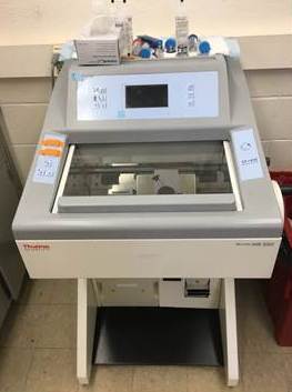 The Department of Physiology's Histology Core contains a ThermoFischer HM 550 Cryostat capable of sectioning tissue at various subzero temperatures. Section thickness can be varied from single microns to several hundred microns. The available Vacutome option keeps the cryostat free of contaminant tissue (important in RNA work) and allows for disposal of unwanted sections. The Vacutome is also useful for reducing curling of tissue for immunohistochemsity or in situ hybridization studies, and is valuable for sectiong tissue for later laser capture microdissection.
The Histology Core has also made available for use a standard brightfield Nikon Eclipse Ci compound microscope with 2X, 10X, 20X, 40X objectives and a Spot Insight Color Camera for viewing and capturing digital images.
 Orientation and prior sign-up is required for use of all equipment and can be arrange through Toni Azar (tazar@med.wayne.edu) or Dr. Mueller (pmueller@med.wayne.edu). Until further notice, equipment is available free of charge (minus own materials) to all Physiology personnel. Costs to those individuals outside the Department will be assessed on a case by case basis by the Chair of Physiology, Dr. J.-P. Jin (jjin@med.wayne.edu).
 - Integrative Cardiovascular Function
Core Director: Donal O'Leary, Ph.D.
- Mass Spectrometry
Core Director: Xuequn Chen, Ph.D.
Potential applications of the Mass Spectrometry (MS) -based proteomic approach
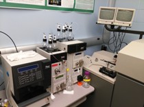 The LC-MS instruments to conduct proteomics analyses include: 1). Dell computer system with Analyst and Bioanalyst software; 2). Dionex nanoLC including Ultimate micropump, Switchos and Famos autosampler; 3). Protana Nanospray source with imaging system; 4). The QSTAR XL Hybrid LC-MS/MS System ideal for iTRAQ-based quantitative proteomics analysis; 5). Remote access to Mascot server in the Proteomics Core in the same building to conduct database searches for mass spec data:
Identification of proteins (organellar proteome, interacting partners etc); Relative quantification of protein abundance with iTRAQ or other methods; Peptide mapping (high sequence coverage); Determination of intact protein molecular weight
- Quantitative PCR
Core Director: Robert Wessells, Ph.D.
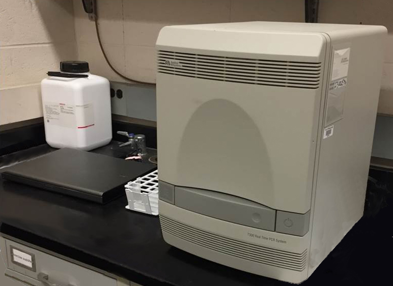
The Quantitative (Q) PCR Facility includes machines for end-point PCR and quantitative RT-PCR. qRT-PCR usable with SYBR-Green or TaqMan detection. 96-wells available. Dedicated computer for machine control and quantitation is located next to qRT-PCR machine. Requires either Opti-clear film cover for plates or Opti-clear capped strips.
Contact e-mail for use is asujkows@med.wayne.edu.